Volumetric Specimen Imaging

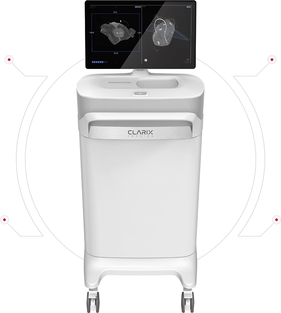

Clarix Imaging VSI-360™
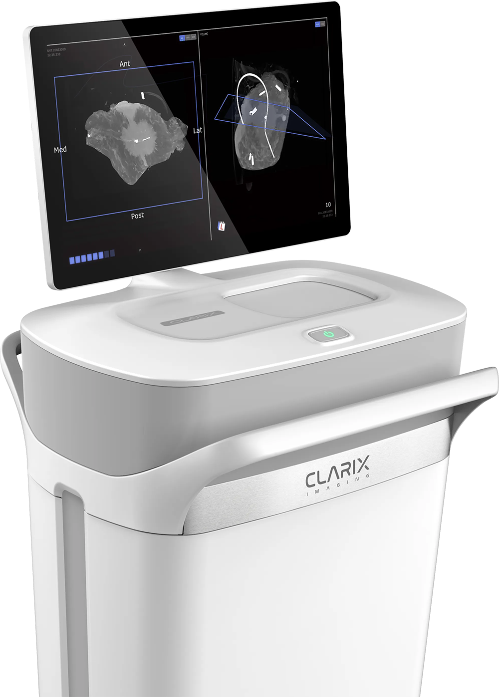
Clarix Imaging’s VSI-360™ generates a true 3D image of the specimen in real-time, and its advanced software tools allow users to visualize the tumor in thin image slices without overlapping tissues obscuring the margins.
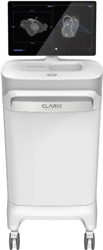
Clarix Imaging VSI-360™
Clarix Imaging’s VSI-360™ generates a true 3D image of the specimen in real-time, and its advanced software tools allow users to visualize the tumor in thin image slices without overlapping tissues obscuring the margins.

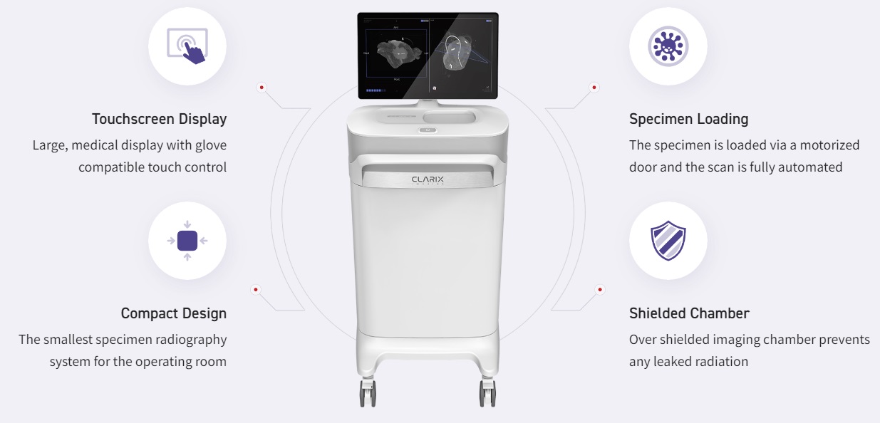
Touchscreen Display
Large, medical display with glove compatible touch control

Intuitive
Minimal training required to operate device and read images

Specimen Loading
The specimen is loaded via a motorized door and the scan is fully automated

Shielded Chamber
Over shielded imaging chamber prevents any leaked radiation
WHY CHOOSE VSI-360TM?
Unique Benefits that Put VSI at a Different Level

Clear
Produces true 3D images with unprecedented clarity

Accurate
Intuitive
Minimal training required to operate device and read images

Convenient
Fits seamlessly into the current OR workflow
A recently published study in the Annals of Surgical Oncology demonstrated 91-94% sensitivity and 81-85% specificity in correlating VSI results to post-surgical pathology1. A recent meta-analysis found that 2D specimen radiography, the current gold standard, has a sensitivity of ~532.
References:
- Kulkarni, Swati A et al. “High-Resolution Full-3D Specimen Imaging for Lumpectomy Margin Assessment in Breast Cancer.” Annals of surgical oncology vol. 28,10 (2021): 5513-5524.
- St John, Edward Robert et al. “Diagnostic Accuracy of Intraoperative Techniques for Margin Assessment in Breast Cancer Surgery: A Meta-analysis.” Annals of surgery vol. 265,2 (2017): 300-310.
WHY CHOOSE VSI-360TM?
Unique Benefits that Put VSI
at a Different Level
Clear
Produces true 3D images with unprecedented clarity
Accurate
Tracks specimen orientation with high precision
Intuitive
Minimal training required to operate device and read images
Convenient
Fits seamlessly into the current OR workflow
A recently published study in the Annals of Surgical Oncology demonstrated 91-94% sensitivity and 81-85% specificity in correlating VSI results to post-surgical pathology1. A recent meta-analysis found that 2D specimen radiography, the current gold standard, has a sensitivity of ~53%2.
References:
- Kulkarni, Swati A et al. “High-Resolution Full-3D Specimen Imaging for Lumpectomy Margin Assessment in Breast Cancer.” Annals of surgical oncology vol. 28,10 (2021): 5513-5524.
- St John, Edward Robert et al. “Diagnostic Accuracy of Intraoperative Techniques for Margin Assessment in Breast Cancer Surgery: A Meta-analysis.” Annals of surgery vol. 265,2 (2017): 300-310.

